
Subscribe to Mental Wealth Academy
>100 subscribers
Subscribe to Mental Wealth Academy
>100 subscribers

The belief that vision happen with our eyes is only partially true. The brain captures light and assembles that light into what we experience as sight. This is how the visual system operates. Photoreceptors in the retina convert light into electrical signals.
What you "see" is already a reconstruction that's shaped by attention, memory, and the brain's predictive models
The raw data our eyes collect is incomplete. Our brain fills the gaps, suppresses irrelevant detail, and prioritizes patterns it recognizes.
Light Is Just Information
Vision begins when light from the sun reflects off objects, and enters the eye through the cornea and pupil. The lens focuses this light onto the retina, a thin sheet of neural tissue lining the back of the eye.
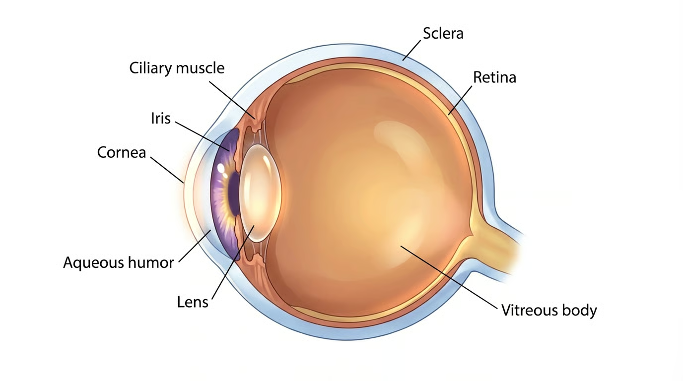
Importantly, the image projected onto the retina is upside down and reversed left to right.

Your brain can't handle it right now so we'll deal with it later.
What you should know is that the light has to be converted into neural signals.
Rods and Cones: Two Ways of Seeing
The retina contains two primary types of photoreceptors:
Rods
Extremely sensitive to light
Enable night vision and peripheral vision
Do not detect color
Dominate the retina outside the fovea
Rods allow you to see in dim environments but sacrifice detail.
Now lets get into the cones….

Cones
Less light-sensitive
Enable color vision and high spatial resolution
Concentrated in the fovea (central vision)
Come in three types (short, medium, long wavelength sensitive)
Cones allow you to read, recognize faces, and see fine detail — but they require bright light.
How Rods and Cones Switch Between Dark and Light
Here’s where vision becomes counterintuitive.
Unlike most neurons, photoreceptors are more active in the dark than in the light.
In Darkness
Photoreceptors continuously release glutamate
Sodium channels remain open
The cell stays relatively depolarized
This high baseline activity signals “no light detected.”
In Light
Photons activate photopigments (rhodopsin in rods, opsins in cones)
A signaling cascade closes sodium channels
The cell hyperpolarizes
Glutamate release decreases
In other words: light reduces neural firing.
Downstream retinal neurons are tuned to detect these changes, allowing the system to encode contrast, edges, and motion rather than absolute brightness.
This mechanism allows the visual system to operate across an enormous range of lighting conditions… from starlight to sunlight.

The Retina Is Already Doing Computation
Before signals ever leave the eye, the retina performs sophisticated processing through layers of bipolar cells, horizontal cells, and amacrine cells.
These circuits:
Enhance contrast
Suppress redundancy
Detect edges and motion
Separate visual features into parallel channels
By the time information reaches the brain, it has already been heavily transformed.
The retina is complex neural network.
From Eye to Brain
Retinal ganglion cells send visual information through the optic nerve. At the optic chiasm, fibers partially cross so that each hemisphere processes the opposite visual field.
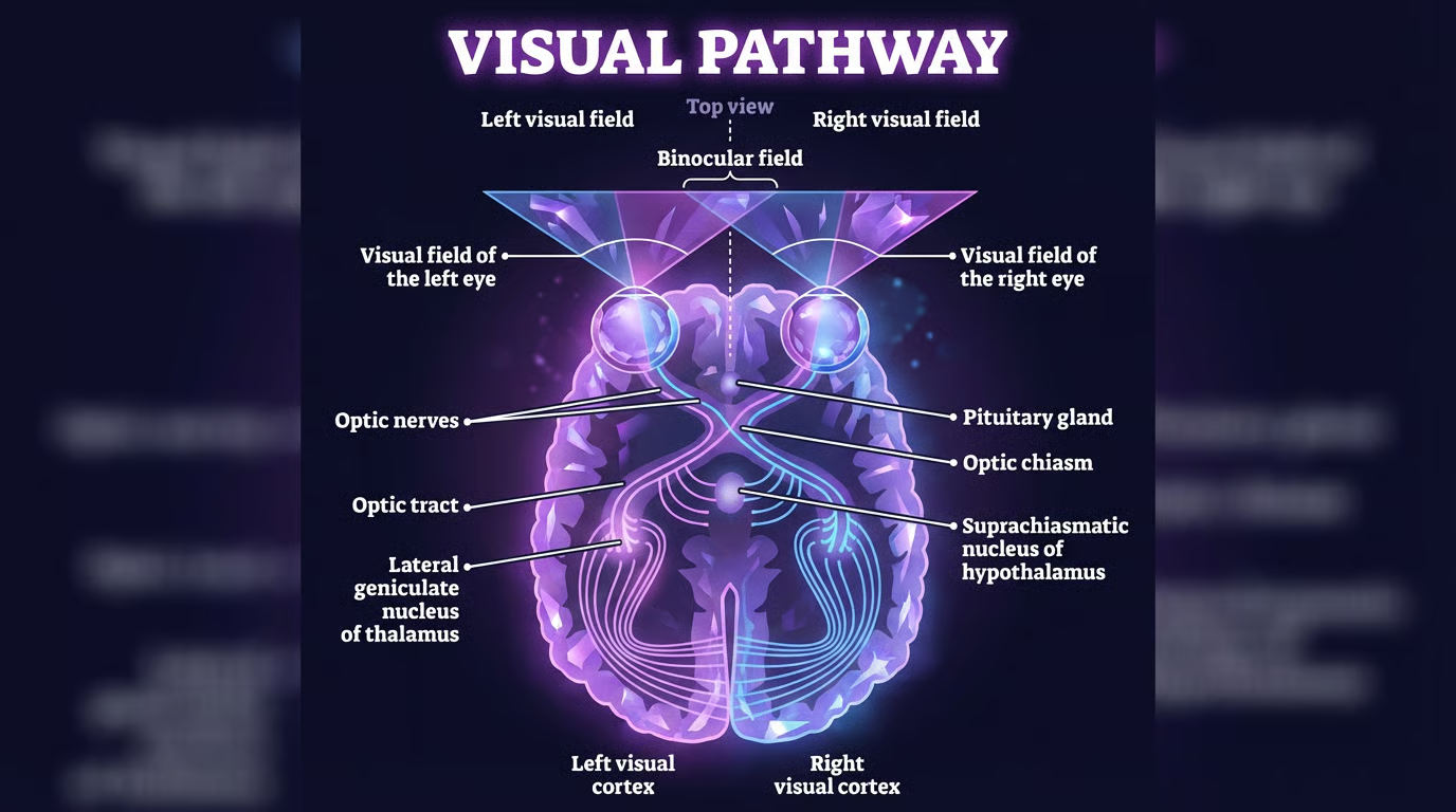
Most signals pass through the lateral geniculate nucleus (LGN) of the thalamus, which:
Regulates signal strength
Integrates attention and arousal
Synchronizes visual input with cortical activity
What you pay attention to influences what gets through.
Seeing the World Upside Down (and Fixing It)
Because of the optics of the eye, the image on your retina is inverted. Yet you don’t experience the world as upside down. This is not because the brain “flips” the image like a photograph. Instead, the brain learns spatial relationships through sensorimotor experience.
From infancy, visual input is continuously paired with:
Movement
Touch
Balance
Proprioception
Over time, the brain builds stable mappings between retinal signals and the external world. Orientation becomes implicit, not calculated.
This is why people wearing inversion goggles (that flip the visual field) can eventually adapt and perceive the world as upright again. Vision is learned how cool is that?
The Visual Cortex Builds Perception
Visual information reaches the primary visual cortex (V1) in the occipital lobe, where neurons respond to:
Orientation
Motion
Spatial frequency
Contrast
From there, processing diverges into two major pathways:
Ventral stream (“what”): object and face recognition
Dorsal stream (“where/how”): motion, depth, spatial interaction

Vision emerges from coordinated activity across populations of neurons.
Vision Is a Prediction
Your brain constantly predicts what it expects to see and updates those predictions based on sensory input.
This explains:
Optical illusions
Context-dependent perception
Why attention changes what you notice
Why fatigue degrades vision
You don’t passively receive the world.
You infer it.
Actionable Takeaways: How to Care for Your Visual System
Vision health is brain health.
1. Respect Light Transitions. Allow time for rods and cones to adapt when moving between bright and dark environments. Sudden lighting changes strain retinal and cortical circuits.
2. Reduce Prolonged Near Focus. Sustained close-up work biases cone-heavy foveal processing. Use the 20–20–20 rule to reduce visual fatigue.
3. Protect Circadian Vision. Retinal signals regulate circadian rhythms.
Get morning daylight
Limit bright screens at night
This supports both sleep and visual clarity.
4. Support Retinal Metabolism. The retina is one of the most energy-demanding tissues in the body.
Maintain metabolic health
Prioritize omega-3 fatty acids
Reduce chronic inflammation
5. Blink Intentionally. Screen use suppresses blinking, degrading visual input quality and increasing cortical effort.
Not all vision problems come from the same place. Some are optical, some are ocular, and some are neurological.
When You Need Glasses. Glasses and contacts correct refractive errors, meaning light is not focused cleanly onto the retina.
Nearsightedness (myopia): distance blur
Farsightedness (hyperopia): near strain or blur
Astigmatism: distorted vision
Presbyopia: age-related difficulty focusing up close
In these cases, the brain is healthy but the image it receives is blurry. Glasses sharpen the signal.
When the Eye Is the Problem. Some conditions damage the eye or retina itself, and glasses cannot fix them.
Cataracts: cloudy lens, reduced contrast
Macular degeneration: loss of central detail
Glaucoma: gradual peripheral vision loss
Retinal disease: disrupted photoreceptor function
When the Brain Is the Problem. Vision can fail even with healthy eyes.
Stroke or brain injury
Visual processing disorders
Difficulty recognizing objects or motion
Here, the eyes detect light, but the brain cannot interpret it correctly.
When to Get Checked. Seek evaluation for sudden vision changes, loss of peripheral vision, flashes of light, or vision changes with headaches or neurological symptoms.
I hope you enjoyed learning about the visual system!
With love,
Dr. Azura Plantiff
Selected References
Masland R. H. (2012). The neuronal organization of the retina. Neuron, 76(2), 266–280. https://doi.org/10.1016/j.neuron.2012.10.002
Hubel, D. H., & Wiesel, T. N. (1968). Receptive fields and functional architecture of monkey striate cortex. The Journal of physiology, 195(1), 215–243. https://doi.org/10.1113/jphysiol.1968.sp008455
By Eric R. Kandel, John D. Koester, Sarah H. Mack and Steven A. Siegelbaum (2021). Principles of Neural Science, Sixth Edition, 6th Edition. McGraw-Hill.
Livingstone, M., & Hubel, D. (1988). Segregation of form, color, movement, and depth: anatomy, physiology, and perception. Science (New York, N.Y.), 240(4853), 740–749. https://doi.org/10.1126/science.3283936
Nassi, J. J., & Callaway, E. M. (2009). Parallel processing strategies of the primate visual system. Nature reviews. Neuroscience, 10(5), 360–372. https://doi.org/10.1038/nrn2619
Friston K. (2010). The free-energy principle: a unified brain theory?. Nature reviews. Neuroscience, 11(2), 127–138. https://doi.org/10.1038/nrn2787
The belief that vision happen with our eyes is only partially true. The brain captures light and assembles that light into what we experience as sight. This is how the visual system operates. Photoreceptors in the retina convert light into electrical signals.
What you "see" is already a reconstruction that's shaped by attention, memory, and the brain's predictive models
The raw data our eyes collect is incomplete. Our brain fills the gaps, suppresses irrelevant detail, and prioritizes patterns it recognizes.
Light Is Just Information
Vision begins when light from the sun reflects off objects, and enters the eye through the cornea and pupil. The lens focuses this light onto the retina, a thin sheet of neural tissue lining the back of the eye.
Importantly, the image projected onto the retina is upside down and reversed left to right.
Your brain can't handle it right now so we'll deal with it later.
What you should know is that the light has to be converted into neural signals.
Rods and Cones: Two Ways of Seeing
The retina contains two primary types of photoreceptors:
Rods
Extremely sensitive to light
Enable night vision and peripheral vision
Do not detect color
Dominate the retina outside the fovea
Rods allow you to see in dim environments but sacrifice detail.
Now lets get into the cones….
Cones
Less light-sensitive
Enable color vision and high spatial resolution
Concentrated in the fovea (central vision)
Come in three types (short, medium, long wavelength sensitive)
Cones allow you to read, recognize faces, and see fine detail — but they require bright light.
How Rods and Cones Switch Between Dark and Light
Here’s where vision becomes counterintuitive.
Unlike most neurons, photoreceptors are more active in the dark than in the light.
In Darkness
Photoreceptors continuously release glutamate
Sodium channels remain open
The cell stays relatively depolarized
This high baseline activity signals “no light detected.”
In Light
Photons activate photopigments (rhodopsin in rods, opsins in cones)
A signaling cascade closes sodium channels
The cell hyperpolarizes
Glutamate release decreases
In other words: light reduces neural firing.
Downstream retinal neurons are tuned to detect these changes, allowing the system to encode contrast, edges, and motion rather than absolute brightness.
This mechanism allows the visual system to operate across an enormous range of lighting conditions… from starlight to sunlight.
The Retina Is Already Doing Computation
Before signals ever leave the eye, the retina performs sophisticated processing through layers of bipolar cells, horizontal cells, and amacrine cells.
These circuits:
Enhance contrast
Suppress redundancy
Detect edges and motion
Separate visual features into parallel channels
By the time information reaches the brain, it has already been heavily transformed.
The retina is complex neural network.
From Eye to Brain
Retinal ganglion cells send visual information through the optic nerve. At the optic chiasm, fibers partially cross so that each hemisphere processes the opposite visual field.
Most signals pass through the lateral geniculate nucleus (LGN) of the thalamus, which:
Regulates signal strength
Integrates attention and arousal
Synchronizes visual input with cortical activity
What you pay attention to influences what gets through.
Seeing the World Upside Down (and Fixing It)
Because of the optics of the eye, the image on your retina is inverted. Yet you don’t experience the world as upside down. This is not because the brain “flips” the image like a photograph. Instead, the brain learns spatial relationships through sensorimotor experience.
From infancy, visual input is continuously paired with:
Movement
Touch
Balance
Proprioception
Over time, the brain builds stable mappings between retinal signals and the external world. Orientation becomes implicit, not calculated.
This is why people wearing inversion goggles (that flip the visual field) can eventually adapt and perceive the world as upright again. Vision is learned how cool is that?
The Visual Cortex Builds Perception
Visual information reaches the primary visual cortex (V1) in the occipital lobe, where neurons respond to:
Orientation
Motion
Spatial frequency
Contrast
From there, processing diverges into two major pathways:
Ventral stream (“what”): object and face recognition
Dorsal stream (“where/how”): motion, depth, spatial interaction
Vision emerges from coordinated activity across populations of neurons.
Vision Is a Prediction
Your brain constantly predicts what it expects to see and updates those predictions based on sensory input.
This explains:
Optical illusions
Context-dependent perception
Why attention changes what you notice
Why fatigue degrades vision
You don’t passively receive the world.
You infer it.
Actionable Takeaways: How to Care for Your Visual System
Vision health is brain health.
1. Respect Light Transitions. Allow time for rods and cones to adapt when moving between bright and dark environments. Sudden lighting changes strain retinal and cortical circuits.
2. Reduce Prolonged Near Focus. Sustained close-up work biases cone-heavy foveal processing. Use the 20–20–20 rule to reduce visual fatigue.
3. Protect Circadian Vision. Retinal signals regulate circadian rhythms.
Get morning daylight
Limit bright screens at night
This supports both sleep and visual clarity.
4. Support Retinal Metabolism. The retina is one of the most energy-demanding tissues in the body.
Maintain metabolic health
Prioritize omega-3 fatty acids
Reduce chronic inflammation
5. Blink Intentionally. Screen use suppresses blinking, degrading visual input quality and increasing cortical effort.
Not all vision problems come from the same place. Some are optical, some are ocular, and some are neurological.
When You Need Glasses. Glasses and contacts correct refractive errors, meaning light is not focused cleanly onto the retina.
Nearsightedness (myopia): distance blur
Farsightedness (hyperopia): near strain or blur
Astigmatism: distorted vision
Presbyopia: age-related difficulty focusing up close
In these cases, the brain is healthy but the image it receives is blurry. Glasses sharpen the signal.
When the Eye Is the Problem. Some conditions damage the eye or retina itself, and glasses cannot fix them.
Cataracts: cloudy lens, reduced contrast
Macular degeneration: loss of central detail
Glaucoma: gradual peripheral vision loss
Retinal disease: disrupted photoreceptor function
When the Brain Is the Problem. Vision can fail even with healthy eyes.
Stroke or brain injury
Visual processing disorders
Difficulty recognizing objects or motion
Here, the eyes detect light, but the brain cannot interpret it correctly.
When to Get Checked. Seek evaluation for sudden vision changes, loss of peripheral vision, flashes of light, or vision changes with headaches or neurological symptoms.
I hope you enjoyed learning about the visual system!
With love,
Dr. Azura Plantiff
Selected References
Masland R. H. (2012). The neuronal organization of the retina. Neuron, 76(2), 266–280. https://doi.org/10.1016/j.neuron.2012.10.002
Hubel, D. H., & Wiesel, T. N. (1968). Receptive fields and functional architecture of monkey striate cortex. The Journal of physiology, 195(1), 215–243. https://doi.org/10.1113/jphysiol.1968.sp008455
By Eric R. Kandel, John D. Koester, Sarah H. Mack and Steven A. Siegelbaum (2021). Principles of Neural Science, Sixth Edition, 6th Edition. McGraw-Hill.
Livingstone, M., & Hubel, D. (1988). Segregation of form, color, movement, and depth: anatomy, physiology, and perception. Science (New York, N.Y.), 240(4853), 740–749. https://doi.org/10.1126/science.3283936
Nassi, J. J., & Callaway, E. M. (2009). Parallel processing strategies of the primate visual system. Nature reviews. Neuroscience, 10(5), 360–372. https://doi.org/10.1038/nrn2619
Friston K. (2010). The free-energy principle: a unified brain theory?. Nature reviews. Neuroscience, 11(2), 127–138. https://doi.org/10.1038/nrn2787
Share Dialog
Share Dialog


No comments yet